Head-injury statistics are staggering. Head injury is the leading cause of death among children and young adults in the United States. According to the
American Association of Neurological Surgeons, someone in the United States receives a head injury every 15 seconds. Every five minutes, one of these individuals dies or becomes permanently disabled. Each year 373,000 Americans are hospitalized for severe head injuries. Of these, 99,000 individuals sustain moderate to severe brain injuries resulting in lifelong disabling conditions.
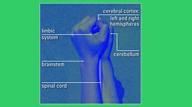
Until recently, what doctors and scientists knew about how the brain works came from observing what stopped working when the brain was injured. Neuroscientists studied thousands of head-trauma victims and methodically mapped specific functions onto various structures of the brain. They found, for instance, that when the frontal lobe of the brain was damaged, patients suffered a loss of motor control. An injury to the temporal lobe of the brain often caused patients to lose the ability to comprehend language and to recognize objects, even though they could still see them. Similarly, researchers found that damage to the limbic system deeper within the brain affected a patient's emotional state.
Obviously, this type of research was painstaking and crude. For starters, head injuries are seldom confined to just one part of the brain but, rather, often involve multiple areas. In addition, different areas of the brain have no visible boundaries between them even when healthy, making the job of correlating structure and function more difficult.
Today, however, neuroscientists can look deep within the human brain without having to wait for a patient with a specific type of head injury to come through the door. They can now observe both properly functioning and improperly functioning brains, including those of children, and make comparisons based on what they see.
One of the latest technologies that allows scientists to do this is called Functional Magnetic Resonance Imaging (fMRI). fMRI scanners detect changes in the oxygen level of the blood flowing through the brain. The level of oxygen in the blood in a given area of the brain is directly related to the level of brain activity in that area. For example, the more oxygen being delivered to the cerebral cortex, an area in the upper middle of the brain, the more active that area is.
Researchers might see this pattern of activity if they asked a patient to read or write. In the brain of a person who has a reading problem, however, they might observe a different pattern of activity. Such observations can improve the accuracy of a diagnosis; in a case like this, a doctor would be able to correctly attribute the patient's reading problem to a problem in the brain, as opposed to poor vision or some other reason.
 Loading Standards

 Print Background Essay