Teachers' Domain - Digital Media for the Classroom and Professional Development
User: Preview

Source: Cold Spring Harbor Laboratory, Dolan DNA Learning Center
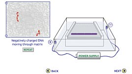
In this interactive activity from the Dolan DNA Learning Center, learn about gel electrophoresis, the standard technique that makes comparative analysis of DNA samples possible. The activity demonstrates, step by step, the process in which DNA fragments are sorted by size as they move through an electrically charged gel matrix.
DNA profiling, also called DNA fingerprinting, is a process that uses DNA to identify individuals. Large portions of DNA are the same for all individuals of a given species—humans included. DNA profiling works because there are sections in which sequences of the four chemical bases (C, G, A, T) vary from individual to individual. By running a procedure called gel electrophoresis, these unique sequences can be used to compare an unidentified DNA sample with samples from known individuals.
During gel electrophoresis, DNA fragments cut from a sample move through a gelatinous medium composed of either agarose—a seaweed extract—or polyacrylamide—a material also used to make soft contact lenses. The gel is cast as a thin slab, with a series of small wells at one end. It then gets placed in a chamber filled with a liquid buffer that can carry an electrical current. Sample DNA fragments are loaded into the empty wells alongside one that gets filled with a marker, sometimes called a DNA ladder. The marker contains DNA fragments whose base sequences and lengths are already known. It is used to help estimate the size of unknown DNA fragments.
When charged molecules are placed in an electric field, they are attracted to poles that carry the opposite charge and repelled from poles that carry the same charge. Phosphate groups in the backbone of DNA molecules give them an overall negative charge. Thus, when an electrical current is applied to the gel, the DNA fragments begin to wiggle through pores in the gel—away from their receptacle wells near the negatively charged end of the slab and toward the opposite, positively charged end of the slab. The moving fragments generate friction, which creates resistance. Because smaller fragments create less friction and therefore less resistance than larger ones, they move faster and farther. In this way, the gel acts as a sieve, separating the molecules by size.
Once the fragments have separated, the current is removed. The gel gets stained with a special dye that binds to DNA and causes the fragments to glow under ultraviolet light. The sample profile from the unknown individual can then be compared to one or more sample profiles from known individuals. If the profile of the unknown individual shows patterns similar to those of a known individual, the samples may have come from the same source. One or two fragments in common cannot ensure a match, but four or five matches can convince scientists beyond a reasonable doubt that the individuals are one and the same.
 Loading Standards
 Teachers' Domain is proud to be a Pathways portal to the National Science Digital Library.
