Teachers' Domain - Digital Media for the Classroom and Professional Development
User: Preview

Source: Produced by WGBH and Digizyme, Inc.


Scientists use a variety of tools to analyze DNA. As this animation produced by WGBH and Digizyme, Inc. shows, gel electrophoresis enables them to determine the size of DNA molecules. Using this technique, together with other tools such as PCR reactions and restriction digestion, scientists can compare the molecular variations of two or more samples to determine such things as the identity of the DNA's source or the presence or absence of a particular gene or DNA fragment.
Gel electrophoresis is a technique used in a wide variety of scientific, criminal, and legal investigations. Although all organisms have a high percentage of genes in common, there are significant genetic differences among members of the same species, even among members of the same family. Scientists use gel electrophoresis to help tease out these differences. As shown in this animation, the technique can also be used in genetic engineering to determine if a sample of bacterial plasmids has been successfully altered to contain a newly inserted gene. But what makes this comparison possible? What makes one molecule of DNA distinguishable from the next?
Gel electrophoresis sorts DNA molecules according to their size and shape. For example, a sample might contain fragments of varying lengths that result from cutting DNA molecules with restriction enzymes, it might contain uncut DNA plasmids, or it might contain a combination of molecules of various sizes and shapes.
To sort these molecules, DNA samples are loaded into wells at one end of the gel, which sits in a bath of buffer solution. The gel is then placed in a special box with electrodes leading from a power source. A negative electrode is attached to one end of the box while a positive electrode is attached to the opposite end. This is important because the DNA molecule itself is negatively charged—a result of the negatively charged phosphate groups that form part of the DNA backbone. When an electrical current is applied to the gel, the DNA molecules move with the current toward the positive end of the gel box. The size and shape of a DNA segment determines the rate at which it moves through the gel.
The gel presents a porous barrier—a matrix—through which DNA segments must pass en route from one end of the gel to the other. The speed with which linear segments of DNA move through the gel when the electrical current is applied is determined by size—the longer the segment, the slower it moves. However, uncut plasmids of identical size can take on different shapes, and this too affects the speed with which they move through the gel. Supercoiled plasmids move much more quickly through the gel than relaxed plasmids because the former are more compact. Finally, when the electrical current is turned off, the molecules in the sample stop moving and are "trapped" at the point in the gel they've reached by that time.
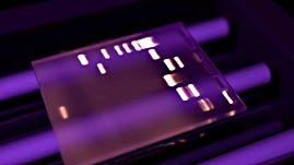
Staining the gel with ethidium bromide after it has run and then exposing it to UV light reveals the location of groups of DNA molecules of similar size and shape. Some lanes of the gel serve as reference points for the test sample. The control sample and the DNA ladder both contain DNA fragments of known size and are used for this purpose. By comparing the bands from the test sample to bands in the ladder and the controls, scientists can assess the relative size and shape of the DNA molecules they contain. In the example shown, a relatively large, slow-moving band in the test lane indicates that the plasmids in the sample do indeed carry a newly inserted gene.
 Loading Standards