Teachers' Domain - Digital Media for the Classroom and Professional Development
User: Preview


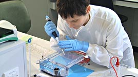
Gel electrophoresis is a powerful technique used to quickly analyze samples for variations in the DNA molecules they contain. While not nearly as precise as decoding an entire genome, gel electrophoresis uses molecular variability to match biological samples to their sources with a great deal of accuracy. However, as this video from the University of Leicester demonstrates, the technique is only as good as the procedures used to conduct the analysis.
Gel electrophoresis is a technique used to detect variations in the size and shape of DNA molecules. The technique is commonly used in forensic DNA analysis and paternity testing to match tissue samples to their sources. It is also used to map genes found on chromosomes to detect mutations that can cause disease. Or, following genetic engineering procedures, gel electrophoresis can be used to determine if DNA molecules have been successfully altered.
There are two main components in a gel electrophoresis procedure. The first is the gel itself, a material made of a complex carbohydrate called agarose. This material, although solid, is also porous and, therefore, allows relatively large molecules such as DNA and RNA to pass through it.
The second main component is the electrophoresis chamber. When electrical current is applied to the chamber, it generates an electrical field, which drives DNA molecules through the gel—this is what is meant by "running the gel." The DNA molecules move through the gel because the negative charges on the molecules' phosphate backbone move away from the negative electrode at one end of the chamber and towards the positive electrode when electrical current is applied.
The speed with which DNA molecules move through the gel, and the distance they travel in a given period of time, depend on the size of the DNA molecules and the agarose concentration of the gel. Small molecules move more quickly and farther than large molecules. All molecules of a given size move more quickly through a gel with a low agarose concentration than through a gel with a high concentration because the molecules experience less resistance. Also, increasing the voltage can increase the rate of movement of the DNA, but up to a limit, since high voltage can also melt the agarose gel.
As the DNA molecules move through the gel while the electric current is applied, there is no way to visibly detect them. For this reason, a dye is included in the sample wells prior to running the gel so that the progress of electrophoresis can be followed even though the movement of the molecules can't be seen.
After electrophoresis is complete, scientists can use a number of chemicals and techniques to make the DNA molecules in the gel visible. One of the most common methods is to stain the gel with a chemical called ethidium bromide (EtBr). EtBr binds directly to the DNA molecule, or intercalates with it, in between the nucleotide bases that make up the rungs of the DNA double helix. More importantly, EtBr fluoresces, or glows, under ultraviolet (UV) light. By viewing the EtBr-stained gel over a UV light source, it is possible to see the DNA bands in the gel.
The DNA bands from unknown or test samples can be compared to DNA bands from known or control samples, including a control sample known as a DNA ladder. DNA ladders are commercially available and are made up of a variety of molecules of known length. When run alongside samples with molecules of unknown length, ladder samples provide a valuable quantitative measure against which the unknowns can be compared in order to assess their size.
Students may benefit from reading the background essay before discussing the following question:
 Loading Standards